
Radiology service
Radiology experts, we provide clear pictures of your health
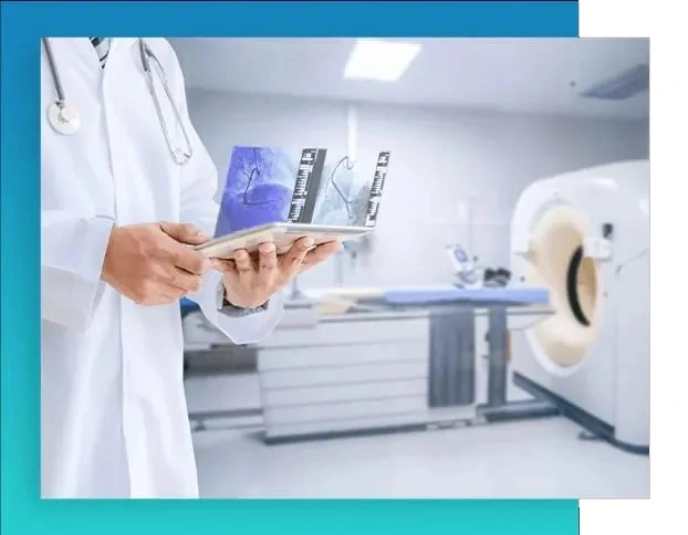
Eshfaa platform provides radiology services with professionalism and high accuracy to its clients.
Radiology service from Eshfaa
Eshfaa platform provides radiology services with professionalism and high accuracy to its clients.
We provide this service in our specialized centers in many governorates. We can also provide radiology services as home visits to cover the whole of Cairo and Giza.
Radiology services at Eshfaa have many advantages:
- It is possible to choose between booking an appointment in the laboratory or a home visit
- Discount offers of up to 25%
- Provides the service of sending results via WhatsApp
- Provide an explanation of the x-ray result to the patient from the medical team, if requested
- A customer service team with a medical background to follow up all the time with the case before and after the tests, respond to its inquiries at any time, and inform it of the required analysis instructions
- Quality of service provided, speed of response and commitment to time.
- The professionalism of the x-ray technicians and the accuracy of the results.
Among the home x-rays available to us, we can offer:
- X ray
- heart drawing
- EEG
- Nerve drawing
- Muscle drawing
- Echocardiogram (ultrasound imaging)
- Ultrasound (abdomen and pelvis, prostate, pregnancy, neck, testicles, thyroid, breast, vaginal)
- doppler

home x-ray
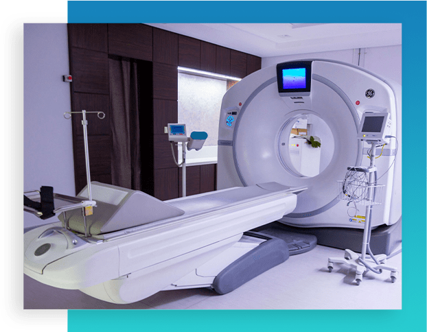
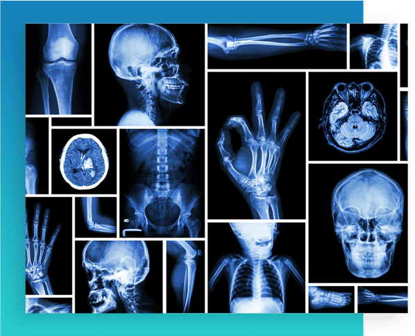
A home X-ray, also known as a home X-ray, is a medical imaging technique that is performed at home.
This technique uses X-rays to produce images of the interior of the body in order to diagnose diseases and assess the condition of internal tissues and structures.
Home x-rays are performed using a portable x-ray machine that is plugged into the mains.
X-rays are directed from the X-ray machine towards the body to be examined, and part of the rays is absorbed by the various tissues in the body. The rays passing through the body are recorded by a special sensor and converted into a digital image showing the internal structures.
Home x-rays have several advantages, including:
- Comfort and Convenience: Having an X-ray at home provides comfort and convenience to patients, as the examination can be performed in a comfortable environment without the need to go to the hospital.
- Time and savings: Getting an x-ray at home saves time and effort associated with going to medical centers.
- Psychological reassurance: Patients can feel more psychological comfort when conducting their medical services at home, where they are in a familiar environment and surrounded by their family members.
- Early detection: Home x-rays enable early detection of many health problems and their speedy treatment
EKG at home
EKG at home
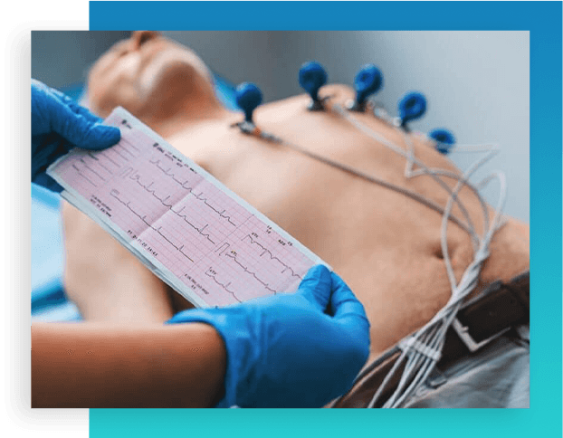
An electrocardiogram, also known as an electrocardiogram, is a medical test used to evaluate the activity of the heart and record the electrical signals that are generated during heartbeats. An electrocardiogram is an important diagnostic tool for detecting heart diseases and arrhythmias.
An EKG is done using a machine called an electrocardiogram (ECG) machine. Electrodes are attached to the patient's body at specific locations such as the chest, arms, and legs. The device records the electrical impulses generated by the heart and converts them into recording waves that reflect the heart's activity.
An electrocardiogram provides important information about the condition and function of the heart, and can identify potential problems such as arrhythmias and abnormal heart formations.
كما يمكن استخدام رسم القلب لتشخيص أعراض مثل ألم الصدر والدوخة وضيق التنفس.
EEG
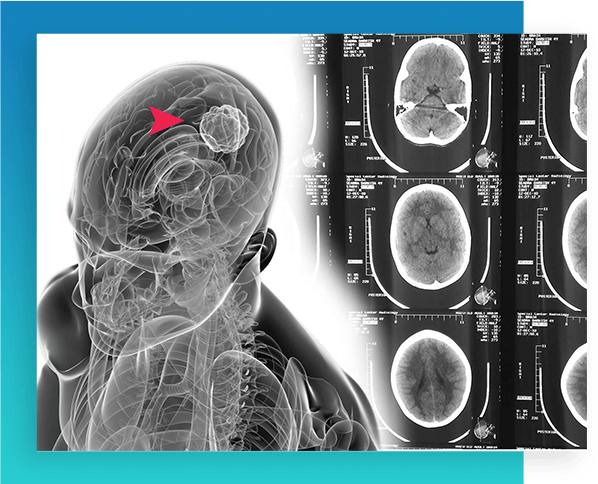
Also known as an electroencephalogram or electroencephalography (EEG), it is a diagnostic test used to record electrical activity in the brain. An EEG aims to evaluate and analyze the electrical activity of the brain and the electrical waves that arise as a result of neural activities.
An EEG is performed by placing small electrodes on the scalp to record electrical signals generated by neurons in the brain.
These electrical signals are recorded by a specialized device called an EEG machine, and converted into a graphic pattern that reflects brain activity.
EEG helps diagnose and monitor many neurological disorders and problems related to the brain, such as epilepsy, neurological spasms, brain tumors, and sleep disorders.
EEGs can also be used to evaluate the brain's response to treatment and to track developments in epilepsy and other brain diseases.
Home EEG
Also known as an electroencephalogram or electroencephalography (EEG), it is a diagnostic test used to record electrical activity in the brain. An EEG aims to evaluate and analyze the electrical activity of the brain and the electrical waves that arise as a result of neural activities.
An EEG is performed by placing small electrodes on the scalp to record electrical signals generated by neurons in the brain.
These electrical signals are recorded by a specialized device called an EEG machine, and converted into a graphic pattern that reflects brain activity.
EEG helps diagnose and monitor many neurological disorders and problems related to the brain, such as epilepsy, neurological spasms, brain tumors, and sleep disorders.
EEGs can also be used to evaluate the brain's response to treatment and to track developments in epilepsy and other brain diseases.
Among the cases that may benefit from a home EEG procedure:
- Patients with mobility difficulties or special needs that prevent them from visiting the hospital.
- Patients who require periodic monitoring of brain activity at home to monitor the effect of treatment or the progression of their condition.
- Young children who find it difficult to remain calm and cooperative in the neurology lab.
- Patients who want to monitor their brain activity during their daily activities in a familiar environment such as home. Young children who find it difficult to remain calm and cooperative in a neurology lab.
However, it must be kept in mind that home EEG may not be available in all cases and may require prior evaluation by the specialist doctor to determine whether this procedure is appropriate and feasible for the patient and his health condition.

Nerve drawing
Nerve drawing
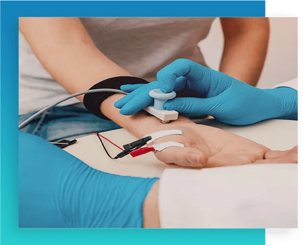
A neurogram is a procedure in which the electrical activity of nerves in the body is recorded. This is done by placing electrodes on the skin or inside the body to record nerve signals. A neurogram is used to diagnose neurological disorders and assess the health and function of nerves.
The electrical signals are recorded using a device called a neurograph or electromyogram. These signals are analyzed to assess the integrity of the nerves and to detect any abnormal changes in nerve activity.
Some common applications of neurography include evaluation of peripheral nerves such as the ulnar nerve, auditory nerve, optic nerve, and sympathetic nerve. It can also be used to diagnose muscle disorders such as muscle spasms, muscle tension, and muscle paralysis.
It is important that the neurogram is performed by physicians who specialize in neurology, as they use the recorded results to determine the correct diagnosis and develop an appropriate treatment plan.
Echocardiogram (ultrasound imaging)
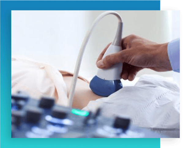
An ultrasound is a procedure that uses sound waves to produce internal images of the tissues and organs in the body. Echocardiography is one of the most common and useful techniques in medical diagnosis.
The echocardiogram is performed using a portable device that contains a transducer that sends out sound waves and records the resulting images. A gel is applied to the skin and the transducer is directed towards the desired area for imaging.
The sound waves are reflected as they hit tissues and organs within the body and captured by the transducer to create 2D or 3D images.
Echocardiography is used to diagnose many conditions and diseases such as heart abnormalities, high blood pressure, thyroid diseases, liver diseases, cancerous tumors, early diagnosis of pregnancy and follow-up of fetal development.
It is a harmless technique and can be done easily and comfortably in a doctor's office.
Interpretation of echo results depends on the expertise of physicians who specialize in ultrasound imaging. Early diagnosis made by echocardiography can help improve treatment outcomes and better guide healthcare.

doppler
Doppler is a procedure used in medical diagnosis to evaluate the movement of blood in blood vessels and internal organs.
Doppler relies on the effect of changing the frequency of sound waves returning from flowing blood to produce a direct picture of blood flow and measure its speed.
Doppler imaging is done using a Doppler ultrasound machine, where sound waves are directed through the skin and reflected off the tissue and blood flow.
Changes in the frequency of the backwaves are analyzed to estimate the speed and direction of blood flow.
Doppler is commonly used to evaluate blood flow in arteries and veins, such as the arteries of the heart and the blood vessels of the neck and extremities. It is also used in the assessment of blood flow in internal organs such as the liver, kidneys, and uterus.
Doppler is a valuable tool in diagnosing many medical conditions such as blocked arteries, heart valve disorders, and liver and kidney diseases.
Doppler helps guide proper treatment and assess the long-term effectiveness of treatment.
It is important that the Doppler procedure is performed by physicians who specialize in medical imaging, and that the results are analyzed and interpreted accurately to achieve a correct diagnosis and direct appropriate treatment.